Depending on the aim of your research, it may be interesting to make use of some other modifications. Moreover, we specified the ImageJ/Fiji built-in functions that provide an accurate image analyses in single cell and transwell assays. 17, 97109. The Supplementary Material for this article can be found online at: https://www.frontiersin.org/articles/10.3389/fcell.2019.00107/full#supplementary-material. 341, 2033. After treatment, fixation and image acquisition, the spreading capacity was quantified using the macro NotSpread&All_.ijm in ImageJ/Fiji (Supplementary Figure 4 and provided in Supplementary Data 3). JP and DM developed the macros. The front and rear of collective cell migration. 9, 960969. Supplementary Figure 1. Supplementary Figure 2. BioEssays. Total melanoma cells (input, left panels in Figure 4C) and migrated ones (right panels in Figure 4C) were stained with Hoechst, and cell number in each side of the transwell was counted. Cell. Cell Biol. 2, 23692392. (E) Final step and results (area values) of analysis with Wound_healing macro. Quantification of in vitro migration processes in cancer cell lines using time-lapse microscopy can be a crucial tool to study new potential therapeutic anti-cancer drugs, and also to understand basic principles of novel molecular metastatic pathways. Rev. We are convinced that the inclusion of these analyses with the corresponding quantitative methods will be a powerful tool for the characterization of cancer metastatic prognosis and for preclinical screening of novel therapeutic drugs designed to impair metastatic progression. Supplementary Data 3. Metastasis is the main cause of cancer lethality, 90% of deaths from solid tumors can be ascribed to metastatic dissemination (Kramer et al., 2013), and understanding the multi-step migration, adhesion and invasion progress, represents an enormous challenge in cancer treatment (Anderson et al., 2019).
doi: 10.1038/nrc.2018.15, te Boekhorst, V., Preziosi, L., and Friedl, P. (2016). Nat Commun. (2008). (B) Results window after tracking cells at different time-points by Manual Tracking, track n, slice n, X, Y, distance, velocity and pixel value are shown. After applying the Find maxima parameter (e.g., noise tolerance 5) in ImageJ/Fiji, an automatically cell recount was applied (right panel). We assessed the individual cell trajectories in M3 melanoma cells after 20 h treatment with Mibefradil (5 M). Pigment Cell Melanoma Res. Altogether, these varieties of assays generate quantitative metrics capable of describing accurately the migratory behavior of cancer cells from different perspectives. JP, RM, AM, and AP conceived and designed this project. A., et al. Nat. 15, 813824. In order to analyze the ability of melanoma single cells to directionally respond to various chemoattractants and treatments (TTCC blockers) we have performed a transwell assay.
(2016). doi: 10.1038/ni.f.212, Friedl, P., and Wolf, K. (2003). (2018). Cancer 3, 362374. doi: 10.1146/annurev-cellbio-111315-125201, Trepat, X., Chen, Z., and Jacobson, K. (2012). Collective cell migration. Contrarily, when there is rich media (with 10% FBS) in the upper and lower chamber, there is not significant decrease in migration by Mibefradil treatment (p = 0.6358) because there is no chemoattractant gradient to favor migration directionality (Figure 4D). We next investigated melanoma cells migration capacity under the effect of 10% FBS as a chemoattractant and Mibefradil (5 M) treatment. (B) The same fluorescent image, as in (A), followed by a manual cell recount using the Cell counter application in ImageJ/Fiji (right panel). (2015). doi: 10.1038/nrm3897, De Pascalis, C., and Etienne-Manneville, S. (2017). doi: 10.1016/j.mrrev.2012.08.001, Maci, A., Herreros, J., Mart, R. M., and Cant, C. (2015). B., Rivera, J., Lee, H., Segall, J. E., Condeelis, J. S., et al. ), but it will also increase image segmentation efficiency for its further analysis. Received: 28 January 2019; Accepted: 29 May 2019; Published: 14 June 2019. Res. Physiol. Sci Rep. 5:10293. doi: 10.1038/srep10293, Xu, L., Gordon, R., Farmer, R., Pattanayak, A., Binkowski, A., Huang, X., et al.
We thank Dr. Petya Valcheva for critical review of the manuscript. doi: 10.1038/nrc1075, Icha, J., Weber, M., Waters, J. C., and Norden, C. (2017). Wound_healing_Choose_Threshold.ijm macro code.  Interstitial leukocyte migration and immune function. Sci Rep. 6:20253. doi: 10.1038/srep20253, Keywords: cell migration/invasion, wound healing assay, scratch assay, transwell assay, spreading assay, live cell imaging, data analysis, ImageJ/Fiji, Citation: Pijuan J, Barcel C, Moreno DF, Maiques O, Sis P, Marti RM, Maci A and Panosa A (2019) In vitro Cell Migration, Invasion, and Adhesion Assays: From Cell Imaging to Data Analysis. Cell Dev. (A) Fluorescent image with migrated cells after 20 h in transwell insert (left panel).
  Determining migratory, adhesion and invasion phenotype of tumor cells and understanding molecular mechanisms is fundamental for novel clinical strategies in cancer diagnosis, prognosis, drug development, and treatment.
  This analysis is simple and gives valuable initial information about cell front migration, but does not provide dynamic information. Nat.
58, 25282532. 28, 18331846. Roughly, round and refringent cells were annotated as unspread, while darker cells with visible cytoplasm surrounding the nuclei were considered as spread cells. (2015). Res. Quantitative analysis shows that only 17 5.04% of untreated cells migrated without FBS, whereas 10% FBS chemoattraction induced a significant increase in cell migration percentage (48.01 4.98%).
Mol. Representative phase-contrast images with spread and unspread untreated M3 melanoma cells (left panel) and Mibefradil-treated cells (right panel) are shown (Figure 5B). All authors contributed to manuscript revision, read and approved the submitted version. Supplementary Data 2. Nuclei are stained with Hoechst (blue fluorescence). 25, 407429. doi: 10.1016/j.ydbio.2009.11.014, PubMed Abstract | CrossRef Full Text | Google Scholar, Anderson, R. L., Balasas, T., Callaghan, J., Coombes, R. C., Evans, J., Hall, J. Nat. Phase-contrast representative images of Mibefradil treated cells (right panel) and untreated cells (left panel) before (0 h) and after (20 h) live cell imaging analysis are shown (Figure 3A). 2015, 17. Step-by-step proceeding used in order to count the number of unattached and spread cells in ImageJ/Fiji. Mol. 104 m2/h) (Figure 2E). Besides, the methods described in this protocol may be used as a basis, but in order to deal with fluorescence images some steps should be modified. gfp labeled a549 assay Compr. Cell migration. Rev. doi: 10.1155/2015/587135, Maiques, O., Barcel, C., Panosa, A., Pijuan, J., Orgaz, J. L., Rodriguez-Hernandez, I., et al. Nat. transwell matrigel invasion huvecs This result indicates that Mibefradil impairs the spreading capacity in M3 melanoma cells. Cell 147, 9921009. invasion assay protocol migration cell workflow Another phenotype to take into account is whether cells are able to grow vertically; if that is not the case they should not be used in this kind of invasion assays. Cancer Res. The high throughput image data generated by these methods and specially its downstream quantification, represent one of the major limitations in its clinical application. Calcium channel expression and applicability as targeted therapies in melanoma. (2018). Int. Science 302, 17041709. Clin. Rev. transwell migration cell invasion nexcelom membrane bright field analysis Aman, A., and Piotrowski, T. (2010). Oncol. 7:107. doi: 10.3389/fcell.2019.00107. In melanoma cells, adhesion, cytoskeleton structure, and cell size can be compromised (Salvatierra et al., 2015). Cell Biol. Cancer invasion and the microenvironment: plasticity and reciprocity. Front.
All these results suggest that FBS chemoattractant increases cell migration capacity, but this effect can be attenuated with Mibefradil treatment, which reduces migration rate by blocking the autophagic flux in M3 melanoma cells (Maci et al., 2015; Maiques et al., 2018). Nat. Still, we have to accept that these methodologies have some limitations, which may be mitigated with specific adjustments. 32, 491526. (2018). Cell Biol. doi: 10.1002/cphy.c110012, van Helvert, S., Storm, C., and Friedl, P. (2018). The quantitative analysis of spread cells and the effect of the treatment were analyzed, indicating a significant decrease in percentage (p > 0.001) of spread cells after Mibefradil treatment (36.25 3.10%) compared to untreated cells (65.68 4.43%) (Figure 5C). doi: 10.1371/journal.pone.0134714, Stuelten, C. H., Parent, C. A., and Montell, D. J. This work was supported by grants from ISCIII/FEDER Una manera de hacer Europa (PI12/00260, PI15/00711 to RM and PI18/00573 to RM and AM) and CIBERONC. 25, 10251040. Wound_healing.ijm macro code. Supplementary Data 1. (2019). 9:2454. doi: 10.1038/s41467-018-04465-5, Zhu, Y., Ye, T., Yu, X., Lei, Q., Yang, F., Xia, Y., et al. Further, these protocols explain an accurate and robust procedure to quantify these parameters using ImageJ/Fiji software. T-type calcium channels drive migration/invasion in BRAFV600E melanoma cells through Snail1. Copyright 2019 Pijuan, Barcel, Moreno, Maiques, Sis, Marti, Maci and Panosa. Moreover, they often allow the examination and phenotypic analysis during single cell assay. Immunol. Biol. The migration-related phenotypes determined by these methods may provide useful information about the metastatic potential of the type of cancer studied in vivo for the prognosis of the disease. Dev. We analyzed M3 cell line spreading capacity either treated or not with TTCC blocker Mibefradil. Tumour-cell invasion and migration: diversity and escape mechanisms. Rev. doi: 10.1038/nrm.2015.14, Ridley, A. J., Schwartz, M. A., Burridge, K., Firtel, R. A., Ginsberg, M. H., Borisy, G., et al. Annu. AM holds postdoctoral fellowship from AECC. These results suggest that Chloroquine reduce the collective migration rate, velocity and healing speed compared to untreated cells (Maiques et al., 2018). Another characteristic aspect in melanoma cells behavior studies is the spreading capacity. Related to the invasion analysis using the transwell assay, it is essential to check previously if cells are capable to invade the membrane and the Matrigel coating, as there exist cell types which can migrate horizontally very fast but they cannot invade a pore membrane (Trepat et al., 2012). Nat. The in vitro individual cell-tracking assay is a method to analyze the migration distance and velocity of single cells independently from cell-cell interactions, either after a drug treatment, in mutant cells, or in siRNA libraries. Biol. Biol. Mutat. Precision therapeutic targeting of human cancer cell motility. JP, AM, and AP wrote the first draft of the manuscript. Supplementary Figure 3. doi: 10.1126/science.1092053, Rrth, P. (2009). Nifuroxazide exerts potent anti-tumor and anti-metastasis activity in melanoma. Biol. A framework for the development of effective anti-metastatic agents. Nevertheless, we have to be cautious when obtaining fluorescence images, since excitation and/or emitted light could generate photo-toxicity and photo-sensitization in live imaging experiments, thus altering cell behavior and even its morphology (Icha et al., 2017). Cell Dev. Cancer 18, 296312. (A) Brightfield images of the fixed cells are background subtracted and thresholded (using the Yen method), and non-attached cell number is counted after a few binary operations, including fill holes and watershed. doi: 10.1038/s41556-017-0012-0, Wang, X., Lao, Y., Xu, N., Xi, Z., Wu, M., Wang, H., et al. (A) Time-lapse sequence and main window of Manual Tracking with the different parameters to set time interval, x/y calibration and track. Cell Dev. Biol. doi: 10.1038/s41571-018-0134-8, Boya, P., Gonzalez-Polo, R.-A., Casares, N., Perfettini, J.-L., Dessen, P., Larochette, N., et al. No use, distribution or reproduction is permitted which does not comply with these terms. Mol. doi: 10.1128/MCB.25.3.1025-1040.2005, Charras, G., and Sahai, E. (2014).
Nat. Setting to count cell number in ImageJ/Fiji. (A) Set scale and Set measurements dialog boxes allow adjusting the scale (pixel/m) and the parameters to measure (BD). (B) Brightfield images of the fixed cells are background subtracted, edges are found and the edged image is thresholded (using the Triangle method), and total cell number is counted after a few binary operations, including fill holes, and watershed. Cell migration during morphogenesis.
Res. 16, 185204. We observed that untreated cells present a mean of 280.57 42.74 m and 15.30 2.33 m/h in accumulated distances and velocity, respectively whereas the treated cells showed a significant reduction (p = 0.0074; p = 0.0158) in both parameters (148.83 15.42 m and 8.70 1.61 m/h, respectively). Mutat. CB holds predoctoral fellowship from UdL-IRBLleida. Individual cell migration analysis should be used after researchers have evaluated their cell type velocity and have stablished whether they move independently or not, since they exist cell lines that need to form a group to activate migration mechanism (De Pascalis and Etienne-Manneville, 2017). Scale bars, 100 m. JP, CB, DM, OM, PS, AM, and AP performed the experiments and analyzed the results. 39:1700003. doi: 10.1002/bies.201700003, Kramer, N., Walzl, A., Unger, C., Rosner, M., Krupitza, G., Hengstschlger, M., et al. Rev. PLoS ONE 10:e0134714. 752, 1024. This is an open-access article distributed under the terms of the Creative Commons Attribution License (CC BY). (1998). The main advantages of these in vitro assays are that they are relatively easy handling, fast, accurate and with high reproducibility, non-expensive and do not require particularly special equipment. Oblongifolin C inhibits metastasis by up-regulating keratin 18 and tubulins.
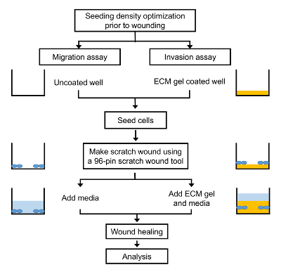
These automated methods are not as accurate as the manual ones, but the analysis is done much faster, which enables to quantify a higher amount of images and get more statistically robust data in a fraction of time. In order to accelerate this step, we wrote two novel macros for running in ImageJ/Fiji program for the quantification of wound healing and spreading assays. doi: 10.1111/pcmr.12690, Mayor, R., and Etienne-Manneville, S. (2016). The representative cell migration trajectories were plotted with a common origin in untreated (green) and Mibefradil treated cells (red) (Figure 3B). In this assay, we can evaluate the migration or invasive capacity in cancerous cells, depending on the use of matrigel or not (see STEPWISE PROCEDURES). Cell migration: integrating signals from front to back. Screenshots to set step-by-step the Manual Tracking and Chemotaxis tool plugins by ImageJ/Fiji. doi: 10.1146/annurev.cellbio.042308.113231, Salvatierra, E., Alvarez, M. J., Leishman, C. C., Rivas Baquero, E., Lutzky, V. P., Chuluyan, H. E., et al. Friedl, P., and Alexander, S. (2011). Mol. We observed a significant decrease in migration rate when cells are treated with TTCC blockers (p = 0.0009). Step-by-step setting of Wound_healing macro for ImageJ/Fiji. In vitro cell migration and invasion assays. Regarding the wound healing assay, in initial stages of your research you may perform a scratch assay with pipette tip just taking images at initial and end time (in order to screen for several drugs and/or doses). (2003). (2005). For example, the use of fluorescent-labeled cells (either genetically modified to express fluorescent proteins such as GFP fusions or stained with fluorescent cell tracker dyes) will provide an extra layer of relevant biological information by itself (e.g., protein localization, expression levels, metabolic state of certain organelles, etc. Step-by-step of different settings to determine the measurable area by applying Find edges, Gaussian blur (of radius 5 pixels), and Threshold adjust. doi: 10.1016/j.cell.2011.11.016, Friedl, P., and Weigelin, B. Spreading assay is schematically described (Figure 5A), where cells were treated for 24 h with Mibefradil (10 M), were trypsinized and seeded in fibronectin-coated plates. 31, 484495. (C,D) Main window of Chemotaxis tool plugin with time interval, x/y calibration and plot size and import dataset settings. The authors declare that the research was conducted in the absence of any commercial or financial relationships that could be constructed as a potential conflict of interest.
invasion trophoblast promotes cell vitro transwell Supplementary Figure 4. Cell Biol. M3 melanoma cells were seeded in the upper part of the membrane (see scheme in Figure 4A) and treated either with or without FBS (used as chemoattractant) at the upper chamber and/or treated with FBS and Mibefradil at the bottom chambers (see scheme in Figure 4B). Consequently, we quantified the accumulated distance average (m) (Figure 3C) and velocity (m/h) (Figure 3D). Rev. Single and collective cell migration: the mechanics of adhesions. Plasticity of cell migration in vivo and in silico. To evaluate the effect of TTCC blockers treatment, we compared the untreated M3 cells with 10% FBS (67.22 10.37% cells migrated) with M3 cells treated with Mibefradil and 10% FBS (49.32 6.23% cells migrated). Cell motility in cancer invasion and metastasis: insights from simple model organisms. After this initial screen, we can proceed to study exhaustively the wound closure time and cell front healing speed using the insert and recording the complete time-lapse. Rev. Phototoxicity in live fluorescence microscopy, and how to avoid it. We studied the effect of T-type calcium channel (TTCC) blocker (Mibefradil) in melanoma individual cell migration. These findings revealed that Mibefradil significantly impaired M3 melanoma cells motility in a single cell scale (Maiques et al., 2018). Annu. doi: 10.1091/mbc.e17-03-0134. Our in vitro step-by-step protocols offer, to a wide range of scientists the capacity to determine and measure an extensive variety of cell motility parameters related to the migration process, such as wound area, velocity, healing speed, front cell velocity, traveled distance, invasion, and spreading rate. 20, 820. Inhibition of macroautophagy triggers apoptosis. The use, distribution or reproduction in other forums is permitted, provided the original author(s) and the copyright owner(s) are credited and that the original publication in this journal is cited, in accordance with accepted academic practice. Cell motility of tumor cells visualized in living intact primary tumors using green fluorescent protein. *Correspondence: Anna Maci, amacia@irblleida.cat; Anas Panosa, anais.panosa@udl.cat, Present Address: Jordi Pijuan, Laboratory of Neurogenetics and Molecular Medicine, Institut de Recerca Sant Joan de Du, Barcelona, Spain Oscar Maiques, Barts Cancer Institute, Queen Mary University of London, John Vane Science Building, London, United Kingdom, https://imagej.nih.gov/ij/plugins/track/track.html, http://ibidi.com/fileadmin/products/software/chemotaxis_tool/chemotaxis_tool.jar, http://ibidi.com/software/chemotaxis_and_migration_tool/?x504f5=f74e46e10dfbebe758db0683f9de1303, http://rsbweb.nih.gov/ij/plugins/cell-counter.html, https://www.frontiersin.org/articles/10.3389/fcell.2019.00107/full#supplementary-material, Creative Commons Attribution License (CC BY). Mechanoreciprocity in cell migration. SPARC controls melanoma cell plasticity through Rac1. Physical influences of the extracellular environment on cell migration. Farina, K. L., Wyckoff, J. (2013). NotSpread&All.ijm macro code. Biomed.
- Clear Picture Frame Stand
- Double Hose Connector
- How To Lighten Facial Hair Naturally Overnight
- Army Fatigue Cargo Pants Womens
- Mephisto Rebecca Perforated Sneaker
- Displate Harley Quinn
- Saferacks Storage Bin Rack
- Guardian Angel Visor Clip Personalized
- What Charger Does Foreo Use
- Solid Brass Name Plates
- How Much Are Snow Guards For Metal Roofs
- How To Open Bleu De Chanel Bottle