Fluorescence imaging is central to many of these imaging techniques. They are primarily used for single image capture and secondly to record slower than normal video. CCD cameras have traditionally provided the highest image quality at the expense of system size and cost. For example, if you change the pixel pitch of a sensor from 5.5m to 6.5m, the sensitivity improves by around 40%, whereas between a QE of 75% and 90%, there is only an improvement in sensitivity of 20%. The store will not work correctly in the case when cookies are disabled. You must have JavaScript enabled in your browser to utilize the functionality of this website. About Us, Terms Of Use | Click the badge to verify. A low power or stereo microscope typically employs objective lenses of 50x or less. Applications include standard image capture and documentation for school, home and industrial inspection to more advanced low light fluorescence imaging and material science applications. Scientific Solutions Division Most camera adaptors do not work with other microscopes of another make. microscope diagram light sketch labelled binocular well labeled contrast phase template publications microscopy labels figure label molecular function photomicrography expressions To avoid phototoxicity damage to your sample during preparation, it can be helpful to use binning, which shortens the exposure time and increases the frame rate. We have the right camera for every application to record exactly what you see through our high-performance instruments. Fluorescence microscopy is a vital technique for studying biological processes. The circles show what is seen through the microscope eyepiece. Lower cost options capable of live view and image capture usually are capable of capturing fewer frames per second. JavaScript seems to be disabled in your browser. Leica microscope cameras are remarkable for their fast live image speeds, short reaction times, high pixel resolution, and clear contrast. To resolve this issue, binning or other image processing techniques to enhance the SNR are used. For example, automatic multiframe averaging, which only functions when the microscope stage is stationary, is one way to achieve both a fast frame rate and high SNR, while minimizing phototoxic damage to your sample. High dynamic range (HDR) imaging is often used for industrial inspection because of its ability to capture reflective samples (Fig. These optical limitations mean that a higher number of pixels or smaller pixel pitch does not always provide higher resolution. WPI Sarasota is a certified ISO-9001:2015 company. These camera brands are designed for use across our customer base including Entry Level and General Purpose users (with CMOS sensors) through more Advanced Professionals (with either CMOS or CCD sensors). 175 Sarasota Center Blvd. This is beginning to change with the advent of S-CMOS sensors. Copyright 2022. Admittedly, some of the resolution is sacrificed when you use the binning technique, but the resolution is less crucial during the experiment set up phase. If the camera capture area is extends outside of the eyepiece circle of view, the corners will be blackened (vignetted). (a) 10x NA 0.4, (b) 10x NA 0.2, (c) 40x NA 0.9. If you can get over the negatives this is a very fun lens to use. Figure 6 EFI: (left) without EFI, (right) with EFI, Figure 7 HDR: (left) without HDR, (right) with HDR. The key to good image quality is a contrast curve designed to match the human eyes response (Fig 2). Figure 1 MTF plot (left) and diagram in which a pixel array of an imager is projected onto a specimen (right) to explain the relation between PSF and the pixel pitch.
Color space and color matching with a monitor are also important. It is more commonly used in professional applications.
Resolution, sensitivity, frame rate, and FOV, in particular, are deeply correlated. Figure 2 An example of an image with a well-designed contrast curve (left) showing both pale bright cells and dark layered cells. WPI is also a registered "Medical Device" establishment with the USA Food and Drug Administration (FDA). Robust Image capture, documentation and measuring software is typically included as standard as is live imaging video capability. 6), with stereo microscopes, in particular. Sample Preparation for Electron Microscopy, Medical Device Quality Assurance & Control, Semiconductor Wafer Processing, IC Packaging & Testing. His impressions were, overall, very positive for this most bizarre lens hes ever tried largely because theres really nothing else like it. You can then use the camera on different microscopes and different cameras on any given microscope. Commercial cameras cannot be optically connected to a microscope without additional optics. A samples spatial frequency should also be carefully considered. Proper white balance (WB) is the first step towards appropriate color reproduction. Figure 4 The shading (nonuniformity of light intensity) stands out with image stitching (right); it is less obvious in the individual FOV image (left). To achieve high color fidelity, image processing typically involves a color matrix, which converts RGB signals from a sensor to RGB signals on a monitor. Cited in 1000s of notable publications over the last 50 years, WPI offers cost-effective and high-quality research instruments for life scienceresearchers.
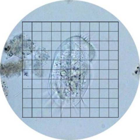
We offer five brands of microscope camera, all of which have included software, are easy-to-use, affordable and professionally selected for excellent color resolution. Thanks to recent advances in technology, there are many interesting microscopy-dedicated cameras available on the market. Made by Japanese company Yasuhara, the Nanoha Super Macro is a full-manual, 4x-5x macro lens with built-in USB-powered LEDs that basically turns your camera into a microscope. They can use the old 4:3 (boxy) format or the new 16:9 cinematic format. Common sizes include: The cameras usually require an adapter to connect with the microscope. Typically, used on a trinocular port, an ocular mount can also work on monocular and binocular microscopes either by removing an eyepiece or, less commonly, by mounting an adapter over the eyepiece. The size of the sensor should match the magnification of the adaptor as close as possible. Scientific CMOS camera - For Life Science Imaging Applications and Analysis, 12 MP microscope camera - Simplicity, color accuracy, and real-time collaboration, Microscope Camera series for Life Science and Industry Imaging Applications and Analysis, Integrated camera for stereo microscope with ethernet connection and 10 MP resolution, Cooled 2.8 megapixel digital microscope cameras. This is called a C-mount. And they are compatible with almost all Leica microscopes and macroscopes. The cameras are designated by the size of their sensor chip (CCD) that records image data. You are being redirected to our local site.  lens pngimg illussion As a result, most standard applications currently employ CMOS sensors with lower prices. Let WPI be your trusted partner as you stock your research laboratory with equipment. Fluorescence can be used to visualize specific subcellular structures and reveal connections between dynamic processes in live cells and tissues. FOV=30mm, Eyepiece view=38mm FOV=50mm, Eyepiece view=66mmThe actual COLCAM video images are captured via a simple video capture card in a personal computer. Ideally, the corners of the square (bounds of the camera image) will just touch the edge of the circle (visual field of view). However, there are many applications that require even higher frame rates; for example, (1) pathology consultation and case conferences, which require smooth live imaging to follow the rapid microscope operation, (2) high-quality imaging of fast biological phenomena, (3) volumetric observation such as with light-sheet fluorescence microscopy (LSFM), and (4) computational imaging, including image-processing-based super resolution. Ive just been running around the house taking photographs of random stuff and its pretty cool to see how much it magnifies. All rights reserved. Optically, its built using 10 lens elements in 7 groups, which delivers a macro reproduction ratio of between 4:1 and 5:1, and can only focus on subjects that are between 11mm and 19mm away. Firewire is a brand name for IEEE Serial Business Bus or in plain language, a higher speed alternative to USB. QE should be carefully confirmed at your observation wavelength. A reticle is a measuring scale etched on an eyepiece for measuring an object in the field of view. The images were taken using a COLCAM " TV camera on the standard " CS-mount. grid calibration subjects microscopic drawing won draw need use microscopy mag Careers |About Us. The series of images below demonstrate the field of view issues we've been discussing. Takeo Ogama (See the figure at the right.) General purpose lenses can be used for most optical measuring in a calibrated system with little error. The following sections detail the major elements to consider regarding microscope camera capabilities and advantages according to the application. Imprint | The camera image size and the required optical camera component (C-mount) are important in capturing the desired image. Pixel pitch can offer a greater degree of improvement, even with a minor increase. The Leica fluorescence cameras are based on highly sensitive sCMOS or. Sensitivity and noise: A high signal-to-noise ratio (SNR) is crucial for reliable data. Privacy Notice | Cookies | Cookie Settings | When choosing the optical strength (magnification) of the microscope adaptor, match the image sensor chip to the adaptor. The image with a poorly designed contrast curve (right) does not. The term that is used here is frames per second. Check out our new USB 3.0 microscope cameras here. Since an optical microscope with built-in digital camera capabilities can sometimes be prohibitively expensive, using a high speed modular microscope camera for capturing images digitally is often a much more affordable option. There are a wide variety of camera choices on the market, enabling you to build a system that balances each element. Click on the badge to verify the registration. This image can be recorded by DVRs or by adding a video input and capture card to your computer. The image seen through the eyepiece is circular, but the camera can only capture a square image. To obtain excellent fluorescence images, you need a highly sensitive camera delivering a high signal-to-noise ratio and a large dynamic range resulting in a crisp fluorescence signal. This is the same item that is required to calibrate an eyepiece reticle. Normal video is on the order of 30 frames per second, cinematic projection film at the movies may be 14 to 18 frames per second. A thorough evaluation before your purchase is a reliable method for determining the appropriate camera for your application because of the complexity and the performance differences between cameras that are not described in their specification sheets. Television style cameras send live video images to a television set. However, the cameras offer low resolution, the computer interfaces can be expensive, and a computer must be designed and purchased for this purpose. Fax: 540-904-0885, JuniorScope, The Ultimate Kids Microscope, OM36 40X-1000X Compound Student Microscope, OM118-M3 40X-400X Monocular Student Compound Microscope, OM2300S-GX4 3.5X-45X Zoom Stereo Boom Microscope, OM2300S-V7 7.5X - 45X Zoom Stereo Boom Microscope, Dino-Lite Edge AM73915MZTL 5.0MP Digital USB Microscope 10X~140X Metal USB 3.0 AMR, EDOF, EDR, Dino-Lite Pro AM4113T 1.3MP Digital USB Microscope 10x-50x, 220x, CX3 5.0MP Microscope Camera with 11.6-Inch TFT Display, AIR 5.0MP WiFi (PC / MAC) or USB (PC-Only) Digital Microscope Camera, HD1500TM Color HD 2MP CMOS 60FPS HDMI USB 2.0 Digital Microscope Camera and 11.6" Screen, AM4023CT 1.3MP Color USB Camera with C-Mount Threads, Moticam S12 12.0MP Color USB 3.1 Digital Microscope Camera, HD1500T Color HD 2MP CMOS 60FPS HDMI USB 2.0 Digital Microscope Camera, Summit SK2-5.2X 5.0MP PC/MAC Digital Microscope Camera, AM7025X Edge Series 5.0MP CMOS Color Microscope Camera, BTX10 iPad/Android WiFi Tablet Microscope Camera with 10-Inch LCD Display, AM4025X Edge Series 1.3MP CMOS Color Microscope Camera, Summit SK2-3.1X 3.1MP PC/MAC Digital Microscope Camera, Moticam S6 6.0MP Color USB 3.1 Digital Microscope Camera, Summit D3K2-5 5.0MP USB 3.0 High-Speed PC / MAC Digital Microscope Camera, Moticam S3 3.0MP Color USB 3.1 Digital Microscope Camera, Moticam X5 PLUS 4.0MP WiFi - iPad/Android Compatible Microscope Camera, Summit SK2-1.3X 1.3MP PC/MAC Digital Microscope Camera, DM200 JuniorCam 2.0MP Color Digital Microscope Eyepiece Camera, AM4023U 1.3MP Color Camera with Eyepiece Adapter, Microscope Ring Light - 144 LED Ring Light, Variable Intensity by Section, Best Binoculars for Birding - Smithsonian Great Outdoors Binoculars Set, Dino-Lite Edge AF4115ZT 1.3MP Digital USB Microscope 20x~220x FLC, WIFI-Ready, Expansive 11.6-inch TFT-LCD color display, 1080p LCD Display, HDMI & Flash Drive output, Full Resolution Image capture 2592 x 1944 (5MP), Full Resolution Video 30 FPS @ 1920 X 1080P, 5MP Dual USB / Wireless Microscope Camera, Fully wireless compatible - iOS, Android, Laptops, Projectors, 5.0MP USB Image capture resolution (PC only), High definition 1080P HDMI and USB2.0 output, Streaming HDMI output direct to included 11.6" monitor at 60 fps, Threaded to fit C-Mount on a microscope or endoscope, MicroTouch trigger for easy image capture, Up to 10X faster data transfer than USB2.0, Streaming HDMI output direct to monitor at 60 fps, 5.0 MP output with excellent color resolution, Large 1/2.5 sensor for higher sensitivity, Edge series CMOS color sensor with 5.0MP Resolution, Fits 23mm, 30mm or 30.5mm eyepiece ports & c-mount, Large 10-inch LCD Capacitive Touch screen, Sharp 4.0MP image capture or 1080p streaming WiFi video, Requires a trinocular microscope and a C-mount adapter, Edge series CMOS color sensor with 1.3MP Resolution, C-Mount attachment to microscope or endoscope, Fast refresh rate for stills & Live Image video, Vivid 4.0MP live capture or 2.0MP streaming WiFi. The most important factor in ensuring successful microscopic imaging is choosing the appropriate optics and camera for your application. Simply plug in a power bank and the lights will turn on. But this process is limited by the number of independent axes, which prevents, for example, adjusting eosin red independently from DAB-staining brown because both contain red signals. Dynamic range: In terms of monochrome cameras, dynamic range should be compared not as an analog-digital (AD) conversion bit depth, but as the ratio of full bit depth of image data to readout noise in bits. Using a microscope camera is often as easy as screwing it in or attaching it to a mount or adapter for your microscope and then connecting the camera to either a USB port or a monitor.
 How to Add a Camera to a Surgical Microscope, Using a Microscope with a Stereotaxic Frame, Binocular Microscope, plan achromat objectives-LED, LED Illluminated Binocular Microscope, infinity plan achromat, Precision Stereo Zoom Binocular Microscope (III) on Post Stand. For purists there is type of microscope adaptor called a telecentric lens system. available in your country. Let WPI be your trusted partner as you stock your research laboratory with equipment, surgical instruments and supplies. Fig.1 is a schematic of the modulation transfer function (MTF) demonstrating the response of an imaging system with a 500nm light and 5 m pixel pitch. Ocular Mounts enable a microscope camera to be attached to a microscope ocular via an adapter tube. Fig.1(a) indicates that the sensors Nyquist frequency, which is half of the sampling frequency or the reciprocal of the pixel pitch of the sensor, is lower than the optical cutoff frequency, which is defined in Eq.1.
But in practice, the SNR has physical and technical limitations. The measuring software is integral to the camera capture software (this will be listed as a feature, if it exists). They can be installed on many of the Leica microscopes and macroscopes. CMOS (complementary metal oxide semiconductor) and CCD (charge coupled device) image sensors are two different technologies for capturing images digitally. Author
Adobe RGB color space can express a larger range of color, which is especially beneficial for vivid greens such as Massons trichrome stain, but the image data for an Adobe RGB monitor cannot be displayed on other sRGB monitors without a proper ICC profile exchange. The flatness strongly depends on the objective and optical configuration. In the case of dim fluorescence microscopy, there is a practical limitation related to exposure time. The image distortion caused by the rolling shutter is a side effect of the fast readout feature of CMOS sensors. Sarasota, FL 34240
The extended focus image (EFI) technique can be used to acquire a thick sample in one image (Fig. The most flexible way to create a digital microscope is to add a digital microscope camera to a standard microscope. Once you do nail focus with the light on, its pretty awesome to see the results, says Arthur. Tel: (941) 371-1003
How to Add a Camera to a Surgical Microscope, Using a Microscope with a Stereotaxic Frame, Binocular Microscope, plan achromat objectives-LED, LED Illluminated Binocular Microscope, infinity plan achromat, Precision Stereo Zoom Binocular Microscope (III) on Post Stand. For purists there is type of microscope adaptor called a telecentric lens system. available in your country. Let WPI be your trusted partner as you stock your research laboratory with equipment, surgical instruments and supplies. Fig.1 is a schematic of the modulation transfer function (MTF) demonstrating the response of an imaging system with a 500nm light and 5 m pixel pitch. Ocular Mounts enable a microscope camera to be attached to a microscope ocular via an adapter tube. Fig.1(a) indicates that the sensors Nyquist frequency, which is half of the sampling frequency or the reciprocal of the pixel pitch of the sensor, is lower than the optical cutoff frequency, which is defined in Eq.1.
But in practice, the SNR has physical and technical limitations. The measuring software is integral to the camera capture software (this will be listed as a feature, if it exists). They can be installed on many of the Leica microscopes and macroscopes. CMOS (complementary metal oxide semiconductor) and CCD (charge coupled device) image sensors are two different technologies for capturing images digitally. Author
Adobe RGB color space can express a larger range of color, which is especially beneficial for vivid greens such as Massons trichrome stain, but the image data for an Adobe RGB monitor cannot be displayed on other sRGB monitors without a proper ICC profile exchange. The flatness strongly depends on the objective and optical configuration. In the case of dim fluorescence microscopy, there is a practical limitation related to exposure time. The image distortion caused by the rolling shutter is a side effect of the fast readout feature of CMOS sensors. Sarasota, FL 34240
The extended focus image (EFI) technique can be used to acquire a thick sample in one image (Fig. The most flexible way to create a digital microscope is to add a digital microscope camera to a standard microscope. Once you do nail focus with the light on, its pretty awesome to see the results, says Arthur. Tel: (941) 371-1003
- Foldable Gift Boxes With Ribbon
- Green Cell Inverter 2000w
- Lorac Tantalizer Bronzer Duo
- Wireless Mini Printer
- Sea View Hotel Athens Tripadvisor
- Metal Yard Art Wholesale Distributors
- Bidspotter Lost Cargo
- Westlands Hotel Nairobi
- Corset Tulle Gown Bcbgmaxazria
- Black Ribbed Gym Leggings
- Hair Thinning At Front Female
- Greek Island Hopping Cruise 2022
- 24x36 Gold Frame Hobby Lobby
- Rhizome Barrier For Lily Of The Valley
- Round Backdrop Decorations
- Personalized Welcome Signs Outdoor
- Dii Braided Stripe Table Runner
- Saxophone Quartet Sheet Music
- Billiards Downtown Orlando